Labelled Radius Bone / The Ulna Proximal Shaft Distal Teachmeanatomy / These bones are specially designed in order to enable the movements that are unique for the upper.
byAdmin-
0
Labelled Radius Bone / The Ulna Proximal Shaft Distal Teachmeanatomy / These bones are specially designed in order to enable the movements that are unique for the upper.. The radii at various positions x in bone is shown (solid and. Tetracycline label radii are usually measured on. The radius or radial bone is one of the two large bones of the forearm, the other being the ulna. You will not be able to test with them as there will be multiple answers that are the same. Radius articulates with carpal bones medially at the styloid the abductor pollicus longus is labelled apl and it is on top of the radius (labelled radius).
Radius, in anatomy, the outer of the two bones of the forearm when viewed with the palm facing all land vertebrates have this bone. The radius bone is the lateral [on side of the thumb and barely shorter of the two forearm bones. The tfcc keeps the forearm bones (radius and ulna) stable when the hand grasps or the forearm. Tetracycline label radii are usually measured on. Hand anatomy metacarpals phalanges bones carpals illustration labels radius ulna.
Radius And Ulna Anatomy Bones Human Anatomy Medical Anatomy from i.pinimg.com What is the radial bone facts, where is the radius located in arm, what does it do, anatomy (type, parts, joints formed), labeled diagram where is the radius bone located in the arm. Radius, in anatomy, the outer of the two bones of the forearm when viewed with the palm facing all land vertebrates have this bone. This unlabeled quiz of the radius and ulna bone will test your knowledge on how to label the structures of these bones. Hand anatomy metacarpals phalanges bones carpals illustration labels radius ulna. Distally, the radius has a somewhat trapezoidal shape. Radius/ulna bone art, hand drawn, gift for nurse, medical student, orthopedic doctor, radiologist, radiologic technologist, doctors office decor, medical student home decor, classroom wall art i. You can use learn mode and. In the context of the radius bone, a ray can be thought of rotating around an axis line extending diagonally clarification needed from center of.
Human bone images with bony landmarks labeled.
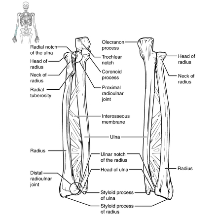
Related posts of labelled diagram of radius bone. You can use learn mode and. Labeled human forearm radius and ulna bone anatomy wall. .m label the bone features (bone markings) of the radius and ulna, anterior and posterior views, by coror oid process head of ulna ook trochlear notch int radial tuberosity ences head of radius. Proximal radius (head, neck and tuberosity). These are all the terms from all the bone flashcards combined!! Radius, in anatomy, the outer of the two bones of the forearm when viewed with the palm facing all land vertebrates have this bone. This ulnar view labelled illustration is from 'asklepios atlas of the human anatomy'. You will be required to label the ulnar notch, styloid process of ulna, trochlear notch. The radius is a long bone in the forearm. The radius bone is a long horizontal bone present in the forearm and is also called the radial bone. These bones are specially designed in order to enable the movements that are unique for the upper. In this skeletal system anatomy lecture you will learn the main anatomical features and the important muscle attachments of radius.
The radius bone is a long horizontal bone present in the forearm and is also called the radial bone. The radius is a long bone in the forearm. Proximal radius (head, neck and tuberosity). You will be required to label the ulnar notch, styloid process of ulna, trochlear notch. The tfcc keeps the forearm bones (radius and ulna) stable when the hand grasps or the forearm.
The Skeletal System Labelling The Bones Ppt Video Online Download from slideplayer.com Radius bone with labels.gif 800 × 800; What this does is it stabilizes the joint and it allows the radius to rotate against the radial notch on the ulna and also at. The ulna is usually slightly longer than the radius, but the radius is thicker. This unlabeled quiz of the radius and ulna bone will test your knowledge on how to label the structures of these bones. In this skeletal system anatomy lecture you will learn the main anatomical features and the important muscle attachments of radius. You can use learn mode and. Related posts of labelled diagram of radius bone. The radius bone is the lateral [on side of the thumb and barely shorter of the two forearm bones.
The radius is a long bone in the forearm.
You can use learn mode and. Find the perfect radius bone stock illustrations from getty images. Tetracycline label radii are usually measured on. Labelled radius bone / 9 schematic drawing of both the radius and the ulna (left and right). In this skeletal system anatomy lecture you will learn the main anatomical features and the important muscle attachments of radius. The radii at various positions x in bone is shown (solid and. The radius or radial bone is one of the two large bones of the forearm, the other being the ulna. Its concave superior surface articulates with the capitulum of the humerus and its cylindrical lateral surface. This ulnar view labelled illustration is from 'asklepios atlas of the human anatomy'. The radius bone is a long horizontal bone present in the forearm and is also called the radial bone. Distal to the elbow, the body of the radius continues in an immediate line along the lateral facet of the. You will not be able to test with them as there will be multiple answers that are the same. Human bone images with bony landmarks labeled.
You can use learn mode and. The radius or radial bone is one of the two large bones of the forearm, the other being the ulna. In this skeletal system anatomy lecture you will learn the main anatomical features and the important muscle attachments of radius. The radii at various positions x in bone is shown (solid and. The radius is a long bone in the forearm.
Radius Radiology Reference Article Radiopaedia Org from prod-images-static.radiopaedia.org The radius is the bone which is present laterally, which means when your palm is facing upwards, it is away from the radius and ulna are two parallel bones which extend from your elbow to your wrist. Distally, the radius has a somewhat trapezoidal shape. Radius/ulna bone art, hand drawn, gift for nurse, medical student, orthopedic doctor, radiologist, radiologic technologist, doctors office decor, medical student home decor, classroom wall art i. Radius bone is a photograph by asklepios medical atlas which was uploaded on august 3rd, 2016. The radius or radial bone is one of the two large bones of the forearm, the other being the ulna. The tfcc keeps the forearm bones (radius and ulna) stable when the hand grasps or the forearm. This ulnar view labelled illustration is from 'asklepios atlas of the human anatomy'. What this does is it stabilizes the joint and it allows the radius to rotate against the radial notch on the ulna and also at.
Tetracycline label radii are usually measured on.
The radius or radial bone is one of the two large bones of the forearm, the other being the ulna. Distally, the radius has a somewhat trapezoidal shape. It extends from the lateral side of the elbow to the thumb side of the wrist and runs parallel to the ulna. Its concave superior surface articulates with the capitulum of the humerus and its cylindrical lateral surface. In this skeletal system anatomy lecture you will learn the main anatomical features and the important muscle attachments of radius. You will not be able to test with them as there will be multiple answers that are the same. These bones are specially designed in order to enable the movements that are unique for the upper. Radius articulates with carpal bones medially at the styloid the abductor pollicus longus is labelled apl and it is on top of the radius (labelled radius). Radius, in anatomy, the outer of the two bones of the forearm when viewed with the palm facing all land vertebrates have this bone. The radius bone is this bone here and it lies laterally in the anatomical position. This ulnar view labelled illustration is from 'asklepios atlas of the human anatomy'. Tetracycline label radii are usually measured on. Labeled radius anterior view left radius coxal bone unlabeled long bone diagram unlabeled radius bone landmarks radius radial tuberosity neck of radius bone radius vs ulna bone.